-
- 抗原
- SNAP Tag
- 宿主
- 兔
- 克隆类型
- 多克隆
- 应用范围
- ELISA, Western Blotting (WB), Scanning Electron Microscopy (SEM)
- 特异性
- Rabbit Anti-SNAP-tag Polyclonal Antibody specifically reacts with fusion proteins containing the SNAP-tag.
- 纯化方法
- Immunoaffinity chromatography
- 免疫原
- Purified recombinant SNAP-tag fusion protein
- 亚型
- IgG
-
- 应用备注
-
Working concentrations for specific applications should be determined by the investigator. The appropriate concentrations may be affected by secondary antibody affinity, antigen concentration, the sensitivity of the method of detection, temperature, the length of the incubations, and other factors. The suitability of this antibody for applications other than those listed below has not been determined. The following concentration ranges are recommended starting points for this product.
ELISA: 0.05-0.2 µg/mL
Western blot: 0.1-1.0 µg/mL
Other applications: user-optimized - 限制
- 仅限研究用
-
- by
- Okeanos Research Laboratory, Department of Biological Sciences, Clemson University
- No.
- #100068
- 日期
- 2016.09.26
- 抗原
- SNAP Tag
- Lot Number
- 13D000621
- Method validated
- Western Blotting
- Positive Control
- E. Coli ER2566 expressing CBD-SNAP
- Negative Control
- E. Coli ER2566 with no CBD-SNAP expression
- Notes
- We confirm that the SNAP Tag antibody ABIN1573927 specifically labels the SNAP-tag in WB in bacterial SNAP-CBD overexpression lysates.
- Primary Antibody
- ABIN1573927
- Secondary Antibody
- anti-Rabbit IgG Alkaline Phosphatase antibody (Sigma-Aldrich, A3687, 060M6006)
- Full Protocol
- Transform E. coli (strain ER2566) with SNAP-CBD expression plasmid (plasmid pYZ205) carrying ampicillin-resistant gene.
- Grow four independent colonies for 12h at 37°C in 25g/L LB (Miller’s) Broth (Growcells, MBPE-1020, lot G14-04) to OD600 0.6.
- Induce SNAP-CBD expression with IPTG (BACHEM 4026913.0005, lot#1049576) at a final concentration of 1mM and growth of the cultures for 4h at 37°C up to OD600 2.5.
- Collect bacterial cells by centrifugation at 3000xg for 10min and freeze pellets at -20°C.
- Select three lysates that show SNAP-CBD expression as positive and one lysate that does not show any SNAP-CBD expression as negative control (see figure).
- Thaw pellets in RIPA lysis buffer (Sigma-Aldrich, R0278, lot SLBL735V) with Protease Inhibitor Cocktail (Sigma-Aldrich, P2714, lot 065M4144V) on ice.
- Determine total protein content of the lysates using Pierce BCA Protein Assay Kit (Thermo Fisher, Reagent A: 23228, lot QE215389; reagent B: 1859078, lot QC216177) compared to a serial dilution of BSA standard (detection range 0-2000µg/ml).
- Denature 25µg and 50µg total protein of the un-/stimulated E. coli culture lysates with loading buffer (Life Technologies, NP0007, lot 1658555) for 5min at 95°C and separate alongside SeeBlue Plus2 Pre-Stained Standard (Thermo Fisher, LC5925, lot 1660839) on a denaturing NuPAGE Novex 4-12% Bis-Tris Gels (Life Technologies, NP0321BOX, lot 15111371) at 185V for 50min.
- Transfer proteins onto nitrocellulose membrane (Amersham Pharmacia Biotech, HybondTM ECLTM Version LRPND/99/5) in a tank blot for 1h at 100V.
- Block membrane with PBST with 1% BSA for 2h at RT.
- Incubate membrane with primary SNAP Tag antibody (antibodies-online, ABIN1573927, lot 13D000621) diluted to 1:500 in 1ml PBST ON at 4°C.
- Wash membrane 3x 30min with PBST with 1% BSA.
- Incubate membrane with secondary anti-Rabbit IgG Alkaline Phosphatase antibody conjugate (Sigma-Aldrich, A3687, 060M6006) diluted to 1:50000 in 5ml PBST with 1% BSA for 2.5h at RT.
- Wash membrane 3x 30min with PBST with 1% BSA.
- AP reactive bands were revealed with 10mL per tablet freshly prepared Sigmafast BCIP/NBT (Sigma-Aldrich, B5655-25TAB, SLBH1944V).
- Experimental Notes
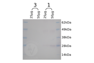
- To validate the specificity of the SNAP Tag antibody ABIN1573927 in Western blot bacterial lysates SNAP-CBD bacterial overexpression lysates were used. We compared the presence of the SNAP-CBD band in lysates that contained SNAP-CBD or not. We found that only in those lysates containing the antigen a protein band of the expected molecular weight was revealed by ABIN1573927.
生效 #100068 (Western Blotting)![成功验证 '独立验证'标志]()
![成功验证 '独立验证'标志]() Validation Images
Validation Images![Western blot analysis of CBD-SNAP tag expression in 25µg and 50µg total protein respectively of bacterial lysates from two different colonies (1 and 3). In colony 1 of approximately 25kDa, the expected size for CBD-SNAP, is revealed where as no band is visible in the negative control culture grown from colony 3.]() Western blot analysis of CBD-SNAP tag expression in 25µg and 50µg total protein respectively of bacterial lysates from two different colonies (1 and 3). In colony 1 of approximately 25kDa, the expected size for CBD-SNAP, is revealed where as no band is visible in the negative control culture grown from colony 3.
Full Methods
Western blot analysis of CBD-SNAP tag expression in 25µg and 50µg total protein respectively of bacterial lysates from two different colonies (1 and 3). In colony 1 of approximately 25kDa, the expected size for CBD-SNAP, is revealed where as no band is visible in the negative control culture grown from colony 3.
Full Methods -
- by
- Okeanos Research Laboratory, Department of Biological Sciences, Clemson University
- No.
- #100069
- 日期
- 2016.09.14
- 抗原
- SNAP Tag
- Lot Number
- 13D000621
- Method validated
- Immunofluorescence
- Positive Control
- Lab stock CBD-SNAP antibody
- Negative Control
- No SNAP-tag antibody
- Notes
- ABIN1573927 specifically labels the SNAP-tag in paraffin embedded oyster visceral mass tissue based on the immunofluorescence staining pattern of CBD-SNAP compared with a CBD-SNAP antibody (lab stock).
- Primary Antibody
- ABIN1573927
- Secondary Antibody
- ABIN101988
- Full Protocol
- Oyster visceral mass tissue is dissected and fixed in 4% paraformaldehyde in seawater overnight.
- Serial dehydration process using an automated ASP300S Enclosed Tissue Processor (Leica Biosystems) as follows:
- 70% ethanol for 45min
- 90% ethanol for 45min
- 90% ethanol for 45min
- 100% ethanol twice for 45min
- xylene twice for 45min
- paraffin wax at 58°C 3 times for 30 min
- Tissue is mounted in a paraffin block and hardened overnight before.
- 8µm tissue sections are retrieved from the block and collected on circular glass cover slips.
- Heat cover slips at 60°C for 1h.
- Deparaffination and rehydration:
- Xylene twice for 15min
- 100% ethanol twice for 10min
- 95% ethanol for 10 min
- 85% ethanol for 10 min
- 70% ethanol for 10 min
- 50% ethanol for 10 min
- 30% ethanol for 10 min
- distilled water for 10 min
- PBS for 10 min
- Wash tissue sections with PBS with 0.05% triton X twice for 30min.
- Permeabilize in PBS with 0.05% triton X overnight.
- Treatment of the tissue sections with 1mg/mL sodium borohydride in PBS three times for 5min to reduce autofluorescence.
- Wash sections in PBS 3 times for 15 min for at RT.
- Block sections in PBST with 1% BSA for 2 hours at RT.
- Incubate sections with CBD-SNAP antibody (lab stock) diluted 1:200 in PBST with 1% BSA overnight at 4°C to detect the location of chitin.
- Wash sections in PBS 3 times for 15min with PBS at RT.
- Additionally, incubate the CBD-SNAP and SNAP-tag double-stained sections with rabbit anti-SNAP antibody (antibodies-online, ABIN1573927, lot 13D000621) diluted 1:200 in PBST with 1% BSA overnight at 4°C.
- Wash sections in PBS 3 times for 15min with PBS at RT.
- Incubate sections with the secondary goat anti-rabbit IgG (heavy & light chain) antibody (FITC) (antibodies-online, ABIN101988, lot 611-1202) diluted 1:400 in PBST with 1% BSA for 2h at °C.
- Wash sections in PBS three times for 15min at RT.
- Counterstain with 0.1µg/mL DAPI in PBS for 15min at RT.
- Wash sections in PBS three times for 15min at RT.
- Mount sections on a microscopic slide using 50% glycerol in PBS.
- Seal cover slips with nail polish.
- Confocal imaging on Leica SPE.
- Visualization of the data performed on LAS 3D software.
- Experimental Notes
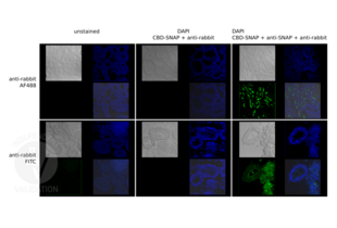
- To validate the specificity of the SNAP Tag antibody ABIN1573927, 8µm paraffin sections of oyster visceral mass were observed in this study. We compared the fluorescence signals with immunofluorescence study. The negative control specimen was always compared with the test specimen or the positive control specimen on the same day, using the same laser power, gain, offset, accumulation/averaging settings on the Leica SPE confocal microscope. Visualization of the data was performed on LAS 3D software, with the same visualization setting to compare signal brightness. We found that the samples incubated with ABIN1573927 had similar fluorescence signals as the positive control CBD-SNAP antibody. Excitation at the same laser wavelength and power did not generate fluorescence in the negative control section, when anti-rabbit FITC secondary antibody was applied in the absence of rabbit produced anti-SNAP antibody
生效 #100069 (Immunofluorescence)![成功验证 '独立验证'标志]()
![成功验证 '独立验证'标志]() Validation Images
Validation Images![Immunofluorescence images of oyster visceral mass tissue, with the specificity of Anti-Rabbit Alexa 488 (top row) and Anti-Rabbit FITC (bottom row) compared. Unstained samples (left column) were compared to test samples (right column) to visualize degree of auto-fluorescence. The absence of anti-SNAP served as the negative control (middle column), it has led to the absence of fluorescence at the same imaging and visualization setting for the green channels, implying the SNAP Tag antibody ABIN1573927 specifically recognizes the SNAP tag.]() Immunofluorescence images of oyster visceral mass tissue, with the specificity of Anti-Rabbit Alexa 488 (top row) and Anti-Rabbit FITC (bottom row) compared. Unstained samples (left column) were compared to test samples (right column) to visualize degree of auto-fluorescence. The absence of anti-SNAP served as the negative control (middle column), it has led to the absence of fluorescence at the same imaging and visualization setting for the green channels, implying the SNAP Tag antibody ABIN1573927 specifically recognizes the SNAP tag.
Full Methods
Immunofluorescence images of oyster visceral mass tissue, with the specificity of Anti-Rabbit Alexa 488 (top row) and Anti-Rabbit FITC (bottom row) compared. Unstained samples (left column) were compared to test samples (right column) to visualize degree of auto-fluorescence. The absence of anti-SNAP served as the negative control (middle column), it has led to the absence of fluorescence at the same imaging and visualization setting for the green channels, implying the SNAP Tag antibody ABIN1573927 specifically recognizes the SNAP tag.
Full Methods -
- by
- Okeanos Research Laboratory, Department of Biological Sciences, Clemson University
- No.
- #100072
- 日期
- 2016.09.14
- 抗原
- SNAP Tag
- Lot Number
- 13D000621
- Method validated
- Electron Microscopy
- Positive Control
- Lab stock CBD-SNAP antibody
- Negative Control
- No SNAP-tag antibody
- Notes
- We validate that the SNAP Tag antibody ABIN1573927 specifically labels the SNAP-tag in SEM-EBSD images of oyster visceral mass tissue.
- Primary Antibody
- ABIN1573927
- Secondary Antibody
- ABIN3042038
- Full Protocol
- Oyster visceral mass tissue is dissected and fixed in 4% paraformaldehyde in seawater overnight.
- Sectiona are is rinsed with 70% ethanol and then transferred to 70% ethanol for about 2 hr at RT before automated dehydration process.
- Serial dehydration process using an automated ASP300S Enclosed Tissue Processor (Leica Biosystems) as follows:
- 70% ethanol for 45min
- 90% ethanol twice for 45min
- 100% ethanol twice for 45min
- xylene twice for 45min
- paraffin wax at 58°C 3 times for 30min
- Tissue is mounted in a paraffin block and hardened overnight.
- 8µm tissue sections are retrieved from the block and collected on circular glass cover slips.
- Heat cover slips at 60°C for 1h.
- Deparaffination and rehydration:
- Xylene twice for 15min
- 100% ethanol twice for 10min
- 95% ethanol for 10min
- 85% ethanol for 10min
- 70% ethanol for 10min
- 50% ethanol for 10min
- 30% ethanol for 10min
- distilled water for 10min
- PBS for 10min
- Wash tissue sections with PBS with 0.05% triton X twice for 30min.
- Permeabilize in PBS with 0.05% triton X overnight.
- Treatment of the tissue sections with 1mg/mL sodium borohydride in PBS three times for 5min to reduce autofluorescence.
- Wash sections in PBS 3 times for 15 min for at RT.
- Block sections in PBST with 1% BSA for 2 hours at RT.
- Incubate sections with CBD-SNAP antibody (lab stock) diluted 1:200 in PBST with 1% BSA overnight at 4°C to detect the location of chitin.
- Wash sections in PBS 3 times for 15min with PBS at RT.
- Additionally, incubate the CBD-SNAP and SNAP-tag double-stained sections with rabbit anti-SNAP antibody (antibodies-online, ABIN1573927, lot 13D000621) diluted 1:200 in PBST with 1% BSA overnight at 4°C.
- Wash sections in PBS 3 times for 15min at RT.
- Incubate sections with the secondary rabbit anti-rabbit IgG (whole molecule) antibody Colloidal Gold (5nm) conjugate (antibodies-online, ABIN3042038) diluted 1:50 in PBST with 1% BSA in the dark overnicht at 4°C.
- Wash sections in PBS three times for 15 min at RT.
- Post fixsections with 1% glutaraldehyde in PBS for 15min at RT.
- Wash sections in distilled water three times for 10min at RT.
- R-Gent SE-EM silver enhancement reagents (Aurion, 2551, lot 60323/2):
- Prepare a slower developer mix with 1:60 ratio of initiator:activator.
- Silver enhancement for 30 min with 1:20 developer: enhancer ratio.
- Stop the reaction by washing with distilled water for 5 min 3 times.
- Graded alcohol series: /li>
- 50% alcohol for 15min
- 70% alcohol for 15min
- 85% alcohol for 15min
- 95% alcohol for 15min
- 100% alcohol for 15min
- Critical point drying of the sections with CO2 transitional fluid for 15min.
- Slowly degas sections at a degassing rate of 100 Pa/min.
- Sections are mounted on the same aluminum stub, coated with carbon and visualized at 20kV with probe current at 40, with basic SEM-EBSD mode on a Hitachi S-3400 Variable Pressure SEM, working distance kept at 10mm. Brightness and contrast settings are kept constant for the both samples.
- Experimental Notes
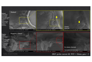
- To validate the specificity of the SNAP Tag antibody ABIN1573927, 8µm paraffin sections of oyster’s visceral mass were observed in this study. We compared the level of silver aggregating ability in the presence and absence of secondary anti-Rabbit IgG (Whole Molecule) Antibody (Colloidal Gold (5nm)) ABIN3042038, using commercially available silver enhancement method and SEM-BSE imaging. We found that the test samples treated with ABIN1573927 showed distinctive silver aggregates which were not present in the negative control.
生效 #100072 (Electron Microscopy)![成功验证 '独立验证'标志]()
![成功验证 '独立验证'标志]() Validation Images
Validation Images![SEM-EBSD images of oyster visceral mass tissue stained with a lab stock CBD-SNAP antibody, anti-SNAP antibody ABIN1573927, and anti-Rabbit IgG (Whole Molecule) secondary antibody (Colloidal Gold (5nm)) and subjected to silver enhancement were imaged in ascending magnification (Top row). As a result clusters of silver aggregates were detected. In the absence of anti-SNAP (Bottom row), the negative control did not show any signs of silver aggregation demonstrating specific CBD-SNAP staining by ABIN1573927. Images in ascending magnification from left to right.]() SEM-EBSD images of oyster visceral mass tissue stained with a lab stock CBD-SNAP antibody, anti-SNAP antibody ABIN1573927, and anti-Rabbit IgG (Whole Molecule) secondary antibody (Colloidal Gold (5nm)) and subjected to silver enhancement were imaged in ascending magnification (Top row). As a result clusters of silver aggregates were detected. In the absence of anti-SNAP (Bottom row), the negative control did not show any signs of silver aggregation demonstrating specific CBD-SNAP staining by ABIN1573927. Images in ascending magnification from left to right.
Full Methods
SEM-EBSD images of oyster visceral mass tissue stained with a lab stock CBD-SNAP antibody, anti-SNAP antibody ABIN1573927, and anti-Rabbit IgG (Whole Molecule) secondary antibody (Colloidal Gold (5nm)) and subjected to silver enhancement were imaged in ascending magnification (Top row). As a result clusters of silver aggregates were detected. In the absence of anti-SNAP (Bottom row), the negative control did not show any signs of silver aggregation demonstrating specific CBD-SNAP staining by ABIN1573927. Images in ascending magnification from left to right.
Full Methods -
- 状态
- Lyophilized
- 溶解方式
- Reconstitute the lyophilized powder with deionized water (or equivalent) to an final concentration of 0.5 mg/ml.
- 缓冲液
- PBS, pH 7.4, containing 0.02 % Sodium azide
- 储存液
- Sodium azide
- 注意事项
- WARNING: Reagents contain sodium azide. Sodium azide is very toxic if ingested or inhaled. Avoid contact with skin, eyes, or clothing. Wear eye or face protection when handling. If skin or eye contact occurs, wash with copious amounts of water. If ingested or inhaled, contact a physician immediately. Sodium azide yields toxic hydrazoic acid under acidic conditions. Dilute azide-containing compounds in running water before discarding to avoid accumulation of potentially explosive deposits in lead or copper plumbing.
- 储存条件
- 4 °C/-20 °C
- 储存方法
- The antibody is stable for 2-3 weeks if stored at 2-8°C. For long term storage, aliquot and store at -20°C or below. Avoid repeated freezing and thawing cycles.
-
-
: "Diffusive tail anchorage determines velocity and force produced by kinesin-14 between crosslinked microtubules." in: Nature communications, Vol. 9, Issue 1, pp. 2214, (2018) (PubMed).
: "Clathrin promotes centrosome integrity in early mitosis through stabilization of centrosomal ch-TOG." in: The Journal of cell biology, Vol. 198, Issue 4, pp. 591-605, (2012) (PubMed).
: "Recombinant human cytoplasmic dynein heavy chain 1 and 2: observation of dynein-2 motor activity in vitro." in: FEBS letters, Vol. 585, Issue 15, pp. 2419-23, (2011) (PubMed).
-
: "Diffusive tail anchorage determines velocity and force produced by kinesin-14 between crosslinked microtubules." in: Nature communications, Vol. 9, Issue 1, pp. 2214, (2018) (PubMed).
-
- 抗原
- SNAP Tag
- 别名
- SNAP-Tag
- 物质类
- Tag
- 背景
- Well-characterized antibodies for epitope tags have been widely used in the study of protein expression in various systems. The SNAP-tag is a self-labeling protein tag that forms a highly stable, covalent thioether bond with fluorophores and other substituted groups on its benzyl guanine substrate. The expressed SNAP-tag fusion proteins can be used in many different assay formats depending upon the labeling substrate employed. Rabbit Anti-SNAP-tag Polyclonal Antibody is developed in rabbit hosts using purified recombinant SNAP-tag fusion protein. This polyclonal antibody is highly purified from rabbit antiserum by immunoaffinity chromatography and is supplied at a concentration of 1 mg/ml in PBS, pH7.4, containing 0.02% Sodium azide.
-
You are here:

 (3 references)
 (3 validations)