Recombinant CRISPR-Cas9 (AA 1-462) 抗体
 (2 validations)
-
- 抗原
- CRISPR-Cas9
- 抗体类型
- Recombinant Antibody
-
抗原表位
- AA 1-462
-
适用
- Staphylococcus aureus
-
宿主
- 小鼠
-
克隆类型
- 单克隆
-
标记
- 非结合性
-
应用范围
- Western Blotting (WB)
- 品牌
- AbFlex®
- 纯化方法
- Protein A Chromatography
- 免疫原
- This antibody was raised against a recombinant protein fragment containing amino acids 1-462 of Staphylococcus aureus Cas9.
- 亚型
- IgG2a
-
anti-CRISPR-Cas9 antibody
适用: Staphylococcus aureus WB, IF 宿主: 兔 Polyclonal unconjugated
anti-CRISPR-Cas9 antibody适用: Streptococcus pyogenes WB, IF, IP, ChIP 宿主: 小鼠 Monoclonal 8C1-F10 unconjugated
anti-CRISPR-Cas9 (C-Term) antibody适用: Streptococcus pyogenes WB, IF, ELISA, FM, MA 宿主: 兔 Polyclonal unconjugated
anti-CRISPR-Cas9 antibody适用: 人 WB 宿主: 小鼠 Monoclonal 1E8 unconjugated
anti-CRISPR-Cas9 (C-Term) antibody (DyLight 549)WB, ELISA, FM 宿主: 兔 Polyclonal DyLight 549
anti-CRISPR-Cas9 (C-Term) antibody (DyLight 800)WB, ELISA, FM 宿主: 兔 Polyclonal DyLight 800
anti-CRISPR-Cas9 (C-Term) antibody (DyLight 680)WB, ELISA, FM 宿主: 兔 Polyclonal DyLight 680
anti-CRISPR-Cas9 (C-Term) antibody (DyLight 488)WB, ELISA, FM 宿主: 兔 Polyclonal DyLight 488
anti-CRISPR-Cas9 (C-Term) antibody (Biotin)适用: Streptococcus pyogenes WB, ELISA, FM 宿主: 兔 Polyclonal Biotin
-
- 应用备注
-
Validated Applications:
WB: 0.5 - 2 µg/ml - 说明
-
AbFlex® antibodies are recombinant antibodies (rAbs) that have been generated using defined DNA sequences to produce highly specific, reproducible antibodies. Each AbFlex antibody contains a 6XHis tag, an avidin tag sequence for enzymatic biotin conjugation using the biotin ligase, BirA, and a sortase recognition motif (LPXTG) to attach a variety of labels directly to the antibody including fluorophores, enzymatic substrates (HRP, AP), peptides, drugs as well as solid supports. AbFlex SaCas9 antibody was expressed as full-length IgG with mouse immunoglobulin heavy and light chains (IgG2a isotype) in mammalian 293 cells.
- 限制
- 仅限研究用
-
- by
- Group Kleinlogel, Department of Physiology, University of Bern, Bern, Switzerland
- No.
- #104486
- 日期
- 2021.04.01
- 抗原
- SaCas9
- Lot Number
- 13018001
- Method validated
- Western Blotting
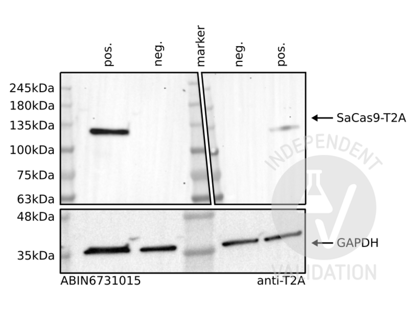
- Positive Control
HEK293 cells transfected with a plasmid expressing SaCas9-T2A-eGFP under the control of the CMV promoter
- Negative Control
- Notes
Passed. ABIN6731015 specifically recognizes saCas9 protein in HEK293 cells.
- Primary Antibody
- ABIN6731015
- Secondary Antibody
- HRP-conjugated goat anti-mouse antibodies (Jackson Immuno Research, 115-035-146)
- Full Protocol
- Grow human embryonic kidney (HEK293) cells (ECACC, 85120602) in DMEM (Sigma, D5671, lot RNB68272) supplemented with 10% Fetal calf Serum (Seraglob, S70500, lot 208/203142), L-Alanyl-L-Glutammine (Merck, Cat. K0302), and Penicillin-Streptomycin (Sigma, P4333, lot 058M4857V) at 37 °C and 5% CO2.
- Plate 0.5x10⁶ cells/ml in 2 ml/well of cells in a 6 well plate.
- Grow cells for 24 h at 37°C and 5% CO2.
- Transfect cells with 1 ug/well of a plasmid expressing SaCas9-T2A-eGFP under the control of the ubiquitous promoter CMV using the calcium phosphate method. For each well, prepare 150 μl 0.5 M CaCl2, add DNA, add 150 μl HBS, incubate for 3 min at RT, and add the whole mix to the cells.
- Change the media after 4-6 h.
- Grow cells for 72 h at 37 °C and 5% CO2.
- Harvest cells with PBS and lyse them on ice for 30 min with 50 µl/well of IP buffer (50 mM TrisHCl pH7-8, 2 mM EGTA, 1 mM EDTA, 0.25% sodium deoxycholate, 1% NP-40 supplemented with protease inhibitors).
- Complete the lysis with a freeze/thaw cycle.
- Determine total protein content of the lysates using Pierce BCA protein assay (Thermo Fisher, 23221).
- Denature 50 μg of total protein for 5 min at 95 °C in 24 μl Laemmli SDS sample buffer and subsequently separate them on a denaturing 4-20% Mini-PROTEAN TGX Stain-Free Gel (Bio-Rad, 456-8094) for 20 min at 100 V and 2 h at 130 V.
- Transfer proteins onto Immobilion-P transfer membrane (Immobilion, IPVH00010) with a Western blotting system for 65 min at 100 V.
- Block the membrane with TBST 5% milk for 1 h at RT.
- Incubate with primary
- mouse anti-SaCas9 antibody (ABIN6731015, lot 13018001) diluted 1:1,000 in TBST 5% milk overnight at 4 °C.
- mouse anti-2A peptide antibody (Novusbio, NBP2 59617, lot A-12) diluted 1:1,000 in TBST 5% milk overnight at 4 °C.
- mouse anti-GAPDH antibody (Fitzgerald, 10R-G09a, lot 2417) diluted 1:4,0000 in TBST 5% milk overnight at 4 °C.
- Wash membrane 3x for 5 min with TBST buffer.
- Incubate with appropriate secondary HRP-conjugated goat anti-mouse antibodies (Jackson Immuno Research, 115-035-146) diluted 1:3,000 in TBST 5% milk for 1 h at RT.
- Wash membrane 3x for 10 min with TBST buffer.
- Reveal protein bands on a ChemiDoc MP imaging system (Bio-Rad, 17001402) using Westar sun ECL Substrate (Cyanagen, XLS063) or Westar ηC2.0 ECL Substrate (Cyanagen, XLS075).
- Experimental Notes
The insertion of a 2A peptide between 2 coding sequences leads to the translation of a unique protein which is then spliced in two proteins by the cell machinery. In this process, the 2A peptide stays bound to the C-terminus of the first protein and it can be used as a tag. In this experiment, the T2A peptide is bound to the saCas9 protein and can be used as a positive control.
The saCas9 antibody ABIN6731015 reveals a protein of the expected molecular weight of antigen in lysates of HEK293 cells. The protein bands are only visible in the positives but not the negative controls. Interestingly, at least in the condition used for this experiment, the saCas9 antibody seemed to provide a stronger signal compared to the T2A antibody.
生效 #104486 (Western Blotting)
 Validation Images
![Western blot analysis of cell lysates from HEK293 cells transfected (pos.) or not transfected (neg.) with a saCas9-T2A-eGFP expression plasmid. Staining was performed using ABIN6731015 (top-left), anti T2A (top-right), or an anti-GAPDH loading control (bottom) antibody.]() Western blot analysis of cell lysates from HEK293 cells transfected (pos.) or not transfected (neg.) with a saCas9-T2A-eGFP expression plasmid. Staining was performed using ABIN6731015 (top-left), anti T2A (top-right), or an anti-GAPDH loading control (bottom) antibody.
Full Methods
Western blot analysis of cell lysates from HEK293 cells transfected (pos.) or not transfected (neg.) with a saCas9-T2A-eGFP expression plasmid. Staining was performed using ABIN6731015 (top-left), anti T2A (top-right), or an anti-GAPDH loading control (bottom) antibody.
Full Methods -
- by
- Group Kleinlogel, Department of Physiology, University of Bern, Bern, Switzerland
- No.
- #104487
- 日期
- 2021.04.01
- 抗原
- SaCas9
- Lot Number
- 13018001
- Method validated
- Immunofluorescence
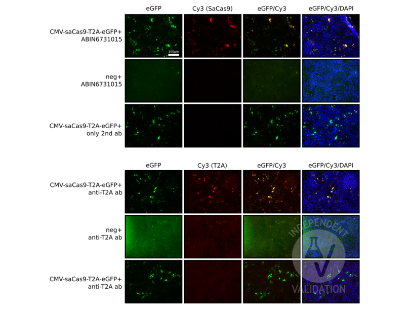
- Positive Control
HEK cells transfected with a plasmid expressing SaCas9-T2A-eGFP
anti-T2A antibody
- Negative Control
- Notes
Passed. ABIN6731015 specifically recognizes SaCas9 in HEK cells expressing SaCas9-T2A-eGFP.
- Primary Antibody
- ABIN6731015
- Secondary Antibody
- NovusBio, NBP2-59627, lot A-12
- Full Protocol
- Grow human embryonic kidney (HEK293) cells (ECACC, 85120602) in DMEM (Sigma, D5671, lot RNB68272) supplemented with 10% Fetal calf Serum (Seraglob, S70500, lot 208/203142), L-Alanyl-L-Glutammine (Merck, Cat. K0302), and Penicillin-Streptomycin (Sigma, P4333, lot 058M4857V) at 37 °C and 5% CO2.
- The day before plating, place a 15mm round glass/well in 24 well plate and incubate overnight with poly L-ornitine (Sigma, P4957).
- Wash with PBS.
- Plate 2x10⁵cells/well in 500 μl in a 24 well plate with round glasses.
- Grow cells for 24 h at 37 °C and 5% CO2.
- Transfect cells with 0.2ug/well of a plasmid expressing human SpCas9-T2A-eGFP under the control of the ubiquitous promoter CMV using the calcium phosphate method. For each well, prepare 30μl 0.25 M CaCl2, add DNA, add 30 μl HBS, incubate for 3 min at RT, and add the whole mix to the cells.
- Change the media after 4-6 h.
- Grow cells for 72 h at 37 °C and 5% CO2.
- Remove the media and wash once with PBS.
- Add 250 μl of 4% PFA and incubate for 7 min.
- Wash 3 times with PBS.
- Add 250 μl of 0.1 M glycine in PBS to block unreacted aldehydes.
- Wash once with PBS.
- Move the coverslips to a microscope slide and circle them with pep-pen.
- Add blocking solution (5% goat serum + 1% BSA Serum in 0.3% Triton-X100 in 1 x TBS) for 60 min at RT. /li>
- Dilute primary
- rabbit anti-saCas9 antibody (antibodies-online, ABIN6731015, lot 13018001) 1:200 in blocking solution.
- mouse anti T2A peptide antibody (NovusBio, NBP2-59627, lot A-12) 1:200 in blocking solution.
- Add 50 µl/well of primary antibody solution.
- Cover the plate with aluminum foil and Incubate overnight at 4 °C in the dark.
- Wash coverslips 3 times with PBS.
- Dilute secondary goat anti mouse Cy3 (Invitrogen, A10521) 1:400 in blocking solution.
- Add 50 µl/coverslips of secondary antibody solution.
- Wash wells 3 times with PBS.
- Add fluorescence mounting medium (DAKO, S3023, lot 10115314) and cover with coverslips. Seal the edges of the coverslips with a clear nail polish.
- Let the nail polish dry and store samples at 4 °C in dark.
- Images were acquired with an Axio VertA1 equipped with an Axiocam 712 Mono and with a 20X objective.
- Experimental Notes
The insertion of a 2A peptide between 2 coding sequences leads to the translation of a unique protein which is then spliced in two proteins by the cell machinery. In this process, the 2A peptide stays bound to the C-terminus of the first protein and it can be used as a tag. In this experiment, the T2A peptide is bound to the SaCas9 protein and it can be used as a positive control. Moreover we expect that cells expressing eGFP should express the saCas9 protein as well.
The SaCas9 antibody ABIN6731015 specifically labels the targeted antigen in HEK293 cells transfected with a plasmid encoding SaCas9-T2A-eGFP. Importantly, cells are always colabeled with eGFP and the antibody.
As a control, the T2A antibody shows a similar pattern.
生效 #104487 (Immunofluorescence)
 Validation Images
![HEK293 cells transfected and not transfected with a plasmid expressing SaCas9-T2A-eGFP and stained with the anti-SaCas9 antibody ABIN6731015 (upper panels) or an anti-T2A antibody (lower panels).]() HEK293 cells transfected and not transfected with a plasmid expressing SaCas9-T2A-eGFP and stained with the anti-SaCas9 antibody ABIN6731015 (upper panels) or an anti-T2A antibody (lower panels).
Full Methods
HEK293 cells transfected and not transfected with a plasmid expressing SaCas9-T2A-eGFP and stained with the anti-SaCas9 antibody ABIN6731015 (upper panels) or an anti-T2A antibody (lower panels).
Full Methods -
- 状态
- Liquid
- 缓冲液
- Purified IgG in 140 mM Hepes, pH 7.5, 70 mM NaCl, 32 mM NaOAc, 0.035 % sodium azide, 30 % glycerol.
- 储存液
- Sodium azide
- 注意事项
- This product contains Sodium azide: a POISONOUS AND HAZARDOUS SUBSTANCE which should be handled by trained staff only.
- 储存条件
- -20 °C
-
- 抗原
- CRISPR-Cas9
- 别名
- Cas9
- 分子量
- 130 kDa
-


