Flotillin 1 抗体 (C-Term)
 (1 validation)
-
- 抗原 See all Flotillin 1 (FLOT1) 抗体
- Flotillin 1 (FLOT1)
-
抗原表位
- C-Term
-
适用
- 人, Cow, 犬, Pig
-
宿主
- 山羊
-
克隆类型
- 多克隆
-
标记
- This Flotillin 1 antibody is un-conjugated
-
应用范围
- Western Blotting (WB), Immunohistochemistry (Paraffin-embedded Sections) (IHC (p))
- 特异性
- This antibody detetcs (FLOT1) at C-term.
- 交叉反应 (详细)
-
Species reactivity (expected):Dog, Pig, Cow.
Species reactivity (tested):Human. - 纯化方法
- Affinity chromatography
- 免疫原
- ptide with sequence C-SISQVNHKPLRTA, from the C Terminus of the protein sequence Genename: FLOT1
- Top Product
- Discover our top product FLOT1 Primary Antibody
-
anti-Flotillin 1 (FLOT1) (C-Term) antibody
Verified FLOT1 适用: 人 WB, ELISA, IHC 宿主: 山羊 Polyclonal unconjugated
anti-Flotillin 1 (FLOT1) (AA 169-251) antibodyFLOT1 适用: 人 WB, ELISA, IHC, IF, IP 宿主: 兔 Polyclonal unconjugated
anti-Flotillin 1 (FLOT1) (AA 128-427) antibodyFLOT1 适用: 人 WB, IHC, IF, IP 宿主: 兔 Polyclonal unconjugated
anti-Flotillin 1 (FLOT1) (C-Term) antibodyKO Validated FLOT1 适用: 人 WB, IP 宿主: 兔 Polyclonal unconjugated
anti-Flotillin 1 (FLOT1) (AA 101-200) antibodyFLOT1 适用: 人, 小鼠, 大鼠 WB, ELISA, IP, IHC (p), IF (cc), IF (p), IHC (fro) 宿主: 兔 Polyclonal unconjugated
anti-Flotillin 1 (FLOT1) (AA 1-427) antibodyFLOT1 适用: 人 WB 宿主: 兔 Polyclonal unconjugated
anti-Flotillin 1 (FLOT1) antibodyFLOT1 适用: 小鼠, 大鼠 WB 宿主: 小鼠 Monoclonal 6C10 unconjugated
anti-Flotillin 1 (FLOT1) (C-Term) antibody (Biotin)Verified FLOT1 适用: 人 WB, ELISA, IHC 宿主: 山羊 Polyclonal Biotin
anti-Flotillin 1 (FLOT1) (AA 219-234), (Middle Region) antibodyFLOT1 适用: 人, 大鼠 WB 宿主: 兔 Polyclonal unconjugated
anti-Flotillin 1 (FLOT1) (AA 104-133), (N-Term) antibodyFLOT1 适用: 人 WB 宿主: 兔 Polyclonal RB40153 unconjugated
-
- 应用备注
-
Peptide ELISA: 1/32000. Western blot: 0,3 - 1,0 1 μg/mL. Approx 48 kDa band observed in H460 lysates (predictedMW of 48 kDa according to NP_005794).
Other applications not tested.
Optimal dilutions are dependent on conditions and should be determined by the user. - 限制
- 仅限研究用
-
- by
- Internal Disease Clinic, Faculty of Veterinary Medicine, University of Zagreb
- No.
- #101253
- 日期
- 2017.05.03
- 抗原
- Flotillin-1
- Lot Number
- Method validated
- Western Blotting
- Positive Control
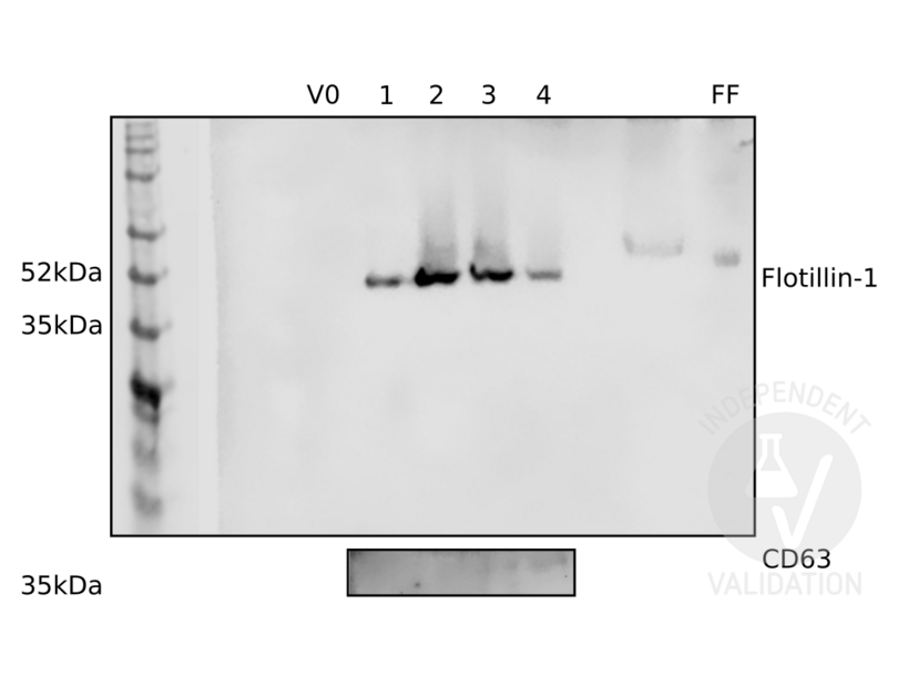
- extracellular vesicles from bovine follicular fluid, verified with CD63 antibody
- Negative Control
- non-enriched fractions
- Notes
- Passed. The flotillin-1 antibody ABIN374222 specifically reveals a band of the expected molecular weight in fractions enriched for exosomes.
- Primary Antibody
- ABIN374222
- Secondary Antibody
- anti-goat-HRP
- Full Protocol
- Isolate extracellular vesicles from 0.5ml follicular fluid from cow obtained from abattoir using the qEV size-exclusion chromatography kit (iZON Science, qEVoriginal Size Exclusion Columns, NC0888135):
- Equilibrate columns with 10ml PBS.
- Apply the follicular fluid.
- Retain the first 3ml of the eluate as void volume negative control (V0).
- Collect the next three 0.5ml fractions (1, 2, 3) which are expected to contain extracellular vesicles.
- Collect next 0.5ml fraction (4) which is expected to contain proteins but not extracellular vesicles from follicular fluid.
- Concentrate fractions 25x using Microcon filters (Millipore, Ultracel YM-30, MRCF0R30) with a 25kDa cut-off.
- Boil all five fractions and 1µl of untreated follicular fluid as positive control in 1x Laemmli SDS sample buffer for 5min.
- Separate all protein in each fraction, 10µg for fraction 2, the richest in protein on a freshly cast 4% acrylamide stacking gel and 10% acrylamide separation SDS-PAGE gel.
- Transfer proteins onto a nitrocellulose membrane (GE Healthcare,Amersham Protran 0.45µm, 10600002) in an electroblotter tank (Biostep, electroblotting module GV100- EBGRM) Western blotting system using 20% methanol-containing transfer buffer for 2h at 150mA at RT.
- Block the membrane with Odyssey blocking buffer (PBS) (Odyssey, 927-400000) for 1h at RT.
- Incubation with primary goat anti-flotillin 1 antibody (antibodies-online, ABIN374222) diluted 1:500 in blocking buffer ON at 4°C.
- Wash membrane 3x 5min with TBST (TBS, 0.1% Tween-20).
- Incubate with secondary anti-goat-HRP antibody diluted 1:5000 in blocking buffer for 1h at RT.
- Wash membrane 3x 5min with TBST.
- Incubate membrane in Luminol for (Santa Cruz Biotechnology Inc., ImmunoCruz western blotting luminol reagent, SC-2048) 5min at RT.
- Reveal chimiuminescent bands using a chemiluminescence detector (LI-COR, Odyssey Fc, OFC-0966) imaging system.
- Strip membranes usingmild stripping protocol:
- Incubate membrane with stripping buffer (1.5% (w/v) glycine, 0.0% (w/v) SDS, 1% (v/v) tween 20, pH2.2) shaking for 2x 10min at RT.
- Wash membrane 2x 10min with PBS.
- Wash membrane2x 5min in TBS.
- Block the membrane with blocking buffer.
- Incubate with primary goat anti-CD63 antibody (antibodies-online, ABIN1440014, lot 0047080912) diluted 1:200 in blocking buffer ON at 4°C.
- Repeat the same steps as for the goat anti-flotillin 1 antibody.
- Experimental Notes
- We observed an albumin band in western blot of some fractions when we used a different secondary (HRP conjugated mouse anti-CD9 antibody) (not shown). Albumin was not observed with ABIN374222.
生效 #101253 (Western Blotting)
 Validation Images
![Different fractions from an enrichment of extracellular vesicles from bovine follicular fluid were probed with antibodies ABIN374222 and ABIN1440014 against exosome markers flotillin-1 (upper panel) and CD63 (lower panel). See protocol for more information. V0: void volume. 1: fraction 1, some exosomes expected. 2: fraction 2, usually the richest in exosomes. 3: third fraction, lower exosome concentration. 4: fourth fraction, contains no or very few exosomes. FF: follicular fluid.]() Different fractions from an enrichment of extracellular vesicles from bovine follicular fluid were probed with antibodies ABIN374222 and ABIN1440014 against exosome markers flotillin-1 (upper panel) and CD63 (lower panel). See protocol for more information. V0: void volume. 1: fraction 1, some exosomes expected. 2: fraction 2, usually the richest in exosomes. 3: third fraction, lower exosome concentration. 4: fourth fraction, contains no or very few exosomes. FF: follicular fluid.
Full Methods
Different fractions from an enrichment of extracellular vesicles from bovine follicular fluid were probed with antibodies ABIN374222 and ABIN1440014 against exosome markers flotillin-1 (upper panel) and CD63 (lower panel). See protocol for more information. V0: void volume. 1: fraction 1, some exosomes expected. 2: fraction 2, usually the richest in exosomes. 3: third fraction, lower exosome concentration. 4: fourth fraction, contains no or very few exosomes. FF: follicular fluid.
Full Methods -
- 浓度
- 0.5 mg/mL
- 缓冲液
- Tris saline, pH ~7.3, 0.02 % Sodium Azide, 0.5 % BSA
- 储存液
- Sodium azide
- 注意事项
- This product contains sodium azide: a POISONOUS AND HAZARDOUS SUBSTANCE which should be handled by trained staff only.
- 注意事项
- Avoid repeated freezing and thawing.
- 储存条件
- 4 °C/-20 °C
- 储存方法
- Store the antibody undiluted at 2-8 °C for one month or (in aliquots) at -20 °C for longer.
-
- 抗原
- Flotillin 1 (FLOT1)
- 别名
- Flotillin-1 / FLOT1 (FLOT1 产品)
- 背景
- Flotillin-1 is a lipid raft-associated protein that has been implicated in various cellular processes: it has been reported to function as a molecular link between lipid rafts of the plasma membrane and a multimeric signaling complex at the actin cytoskeleton, to associate with caveolae , regulating vesicular trafficking and signal transduction, and to have mitogenic properties. Its subcellular localisation seems to be cell-type-specific. It has been found associated with the cell membranes, nucleus, cytoplasm and different cytoplasmic organelles.Synonyms: REG-2, Reg2, Reggie-2
- 基因ID
- 10211
- NCBI登录号
- NP_005794
- UniProt
- O75955
-


