P27 抗体
 (2 validations)
-
- 抗原
- P27
-
适用
- 人, 小鼠, 大鼠, 猴
-
宿主
- 小鼠
-
克隆类型
- 单克隆
-
标记
- 非结合性
-
应用范围
- Western Blotting (WB), Immunohistochemistry (Paraffin-embedded Sections) (IHC (p)), Flow Cytometry (FACS), Immunofluorescence (IF)
- 纯化方法
- Protein G affinity chromatography
- 免疫原
- Mouse recombinant protein (DCS-72.F6) and recombinant human protein (KIP1/769) were used as the immunogen for the p27 antibody cocktail.
- 克隆位点
- DCS-72-F6-KIP1-769
- 亚型
- IgG
-
anti-P27 antibody
适用: 人, 大鼠 IHC 宿主: 兔 Polyclonal unconjugated
anti-P27 (Ser235), (Ser236) antibody适用: 人 WB, ELISA 宿主: 兔 Polyclonal unconjugated
anti-P27 (pThr198) antibody适用: 人 WB, ELISA 宿主: 兔 Polyclonal unconjugated
anti-P27 (pSer10) antibody适用: 人, 小鼠, 大鼠 WB, ELISA 宿主: 兔 Polyclonal unconjugated
anti-P27 (pThr187) antibody适用: 人, 小鼠, 大鼠 WB, ELISA, IHC 宿主: 兔 Polyclonal unconjugated
anti-P27 antibody适用: 人 WB, ELISA, IP 宿主: 兔 Polyclonal unconjugated
anti-P27 antibody适用: 人, 小鼠, 大鼠, 猴 WB, IHC (p), FACS, IF 宿主: 小鼠 Monoclonal DCS-72-F6 unconjugated
anti-P27 (Ser235) antibody适用: 人, 小鼠, 大鼠 WB, ELISA, IHC 宿主: 兔 Polyclonal unconjugated
-
- 应用备注
-
Optimal dilution of the p27 antibody to be determined by the researcher.
1. Staining of formalin-fixed tissues requires boiling tissue sections in 10 mM Citrate buffer, pH 6.0, for 10-20 min followed by cooling at RT for 20 min
2. The prediluted format is supplied in a dropper bottle and is optimized for use in IHC. After epitope retrieval step (if required), drip mAb solution onto the tissue section and incubate at RT for 30 min.\. Flow Cytometry: 0.5-1 μg/million cells in 0.1ml,Immunofluorescence: 0.5-1 μg/mL,Western blot: 0.5-1 μg/mL,Immunohistochemistry (FFPE): 0.25-0.5 μg/mL for 30 min at RT (1),Prediluted format : incubate for 30 min at RT (2) - 限制
- 仅限研究用
-
- by
- Johann-Friedrich-Blumenbach-Institute for Zoology and Anthropology, Department of Developmental Biology, Georg-August-University Göttingen
- No.
- #101901
- 日期
- 2017.12.04
- 抗原
- P27
- Lot Number
- V2438-171009
- Method validated
- Western Blotting
- Positive Control
- NIH/3T3 mouse embryonic fibroblast cells overexpressing mVenus-tagged p27K, starved for 48h
- Negative Control
- Untransfected NIH/3T3 cells, starved for 48h
- Notes
Passed. ABIN3025539 detects the ectopically expressed fusion protein as well as the endogenous protein by immunoblotting. Unspecific cross-reactivity is low.
- Primary Antibody
- ABIN3025539
- Secondary Antibody
- anti-mouse IgG (whole molecule), HRP-linked (Sigma-Aldrich, A9044, lot 034M4761)
- Full Protocol
- Grow NIH/3T3 cells (ATCC, CRL-1658) in DMEM+GlutaMAX (Gibco, 31966-021, Lot 1852045) supplemented with fetal bovine serum (Gibco 270-106) and Pen/Strep (Gibco 15140), at 37°C and 5% CO2 to 70% confluency.
- Transfect cells with a plasmid encoding mVenus-tagged p27K- (kindly provided by Toshio Kitamura, University of Tokyo; Oki et al., 2014) using EndofectinMax (GeneCopoeia) following the manufacturer's instructions.
- Serum starve cells for 48h. Use untransfected NIH/3T3 cells starved for 48h as control.
- Lyse cells in RIPA buffer (10mM PBS pH7.2, 2mM EDTA, 1% NP-40, 1% Triton X-100, protease inhibitors) at 4°C.
- Denature total cellular lysates proteins in 1x SDS-sample buffer and separate proteins on a freshly cast denaturing 10% SDS-PAGE (Laemmli, 1970).
- Transfer proteins onto 0.2µm Protran membrane (GE Healthcare, 10600004, A10043108) with a Western blotting system for 1h at 400A (Towbin et al., 1979).
- Block the membrane in TBST (50mM Tris-HCl, pH7.4, 150mM NaCl, 0.2% Tween 20) containing 5% milk (blocking solution) for 60min at RT.
- Incubate membrane with primary mouse anti-P27 antibody (antibodies-online, ABIN3025539, lot V2438-171009) diluted 1:250 in blocking solution ON at 4°C.
- Incubate membrane with secondary anti-mouse IgG (whole molecule), HRP-linked (Sigma-Aldrich, A9044, lot 034M4761) diluted 1:5000 in TBST for 45min at RT.
- Wash membrane with TBST for 30-45min at RT.
- Reveal protein bands using Clarity Max Western ECL substrate (Bio-Rad, 1705062); image capture via Chemidoc Imaging System (BioRad).
- Experimental Notes
ABIN3025539 reveals a protein band at the expected molecular weight of the mVenus-p27 fusion protein. The antibody does also detect a protein at the expected molecular weight of the endogenous, untagged p27.
生效 #101901 (Western Blotting)
 Validation Images
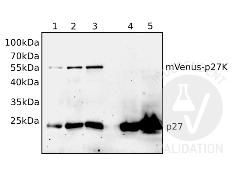
![Different volumes of total cellular lysates of either mVenus-p27K- transfected NIH/3T3 cells (1, 2, 3) or untransfected NIH/3T3 cells (4, 5) were loaded and the blot membrane was incubated with ABIN3025539 followed by incubation with anti-mouse IgG-linked to HRP and chemiluminescence detection. Expected molecular weight of p27 fused to mVenus is approximately 55kDa.]() Different volumes of total cellular lysates of either mVenus-p27K- transfected NIH/3T3 cells (1, 2, 3) or untransfected NIH/3T3 cells (4, 5) were loaded and the blot membrane was incubated with ABIN3025539 followed by incubation with anti-mouse IgG-linked to HRP and chemiluminescence detection. Expected molecular weight of p27 fused to mVenus is approximately 55kDa.
Full Methods
Different volumes of total cellular lysates of either mVenus-p27K- transfected NIH/3T3 cells (1, 2, 3) or untransfected NIH/3T3 cells (4, 5) were loaded and the blot membrane was incubated with ABIN3025539 followed by incubation with anti-mouse IgG-linked to HRP and chemiluminescence detection. Expected molecular weight of p27 fused to mVenus is approximately 55kDa.
Full Methods -
- by
- Johann-Friedrich-Blumenbach-Institute for Zoology and Anthropology, Department of Developmental Biology, Georg-August-University Göttingen
- No.
- #102346
- 日期
- 2017.12.04
- 抗原
- P27
- Lot Number
- V2438-171009
- Method validated
- Immunocytochemistry
- Positive Control
- NIH/3T3 mouse embryonic fibroblast cells overexpressing mVenus-tagged p27K, starved for 48h
- Negative Control
- Untransfected NIH/3T3 cells, starved for 48h
- Notes
Passed. ABIN3025539 detects the ectopically expressed fusion protein by immunocytochemistry. Unspecific cross-reactivity is low.
- Primary Antibody
- ABIN3025539
- Secondary Antibody
- goat anti-mouse IgG (H+L) Alexa Fluor 555 (Invitrogen, A21422, lot 948498)
- Full Protocol
- Grow NIH/3T3 cells (ATCC, CRL-1658) in on cover slips in DMEM, 10% fetal bovine serum (Gibco 270-106), 5% penicillin/streptomycin (Gibco) at 37°C in 5% CO2.
- Transfect cells with a plasmid encoding mVenus-tagged p27K- (kindly provided by Toshio Kitamura, University of Tokyo; Oki et al., 2014) using EndofectinMax (GeneCopoeia) following the manufacturer's instructions.
- Serum starve cells for 48h. Use untransfected NIH/3T3 cells starved for 48h as control.
- Fix cells in 3.7% paraformaldehyde (in PBS) for 15min at 4°C followed by incubation in 0.3% Triton X-100 for 10min.
- Block unspecific binding sites in PBT (phosphate buffered saline (PBS) containing 1% bovine serum albumin, 0.5% Tween-20) for 1h at RT.
- Incubate cells with primary mouse anti-P27 antibody (antibodies-online, ABIN3025539, lot V2438-171009) diluted 1:100 in PBS ON at 4°C.
- Wash cells with TBST (50mM Tris-HCl, pH7.4, 150mM NaCl, 0.1% Tween 20) for 15min.
- Incubate cells with secondary antibody goat anti-mouse IgG (H+L) Alexa Fluor 555 (Invitrogen, A21422, lot 948498) diluted 1:1000 in PBS and DAPI (4’,6-Diamidino-2-phenylindole; Sigma D-9542).
- Image acquisition on Zeiss LSM 510 confocal microscope and processing using Adobe Photoshop 5.0.
- Experimental Notes
生效 #102346 (Immunocytochemistry)
 Validation Images
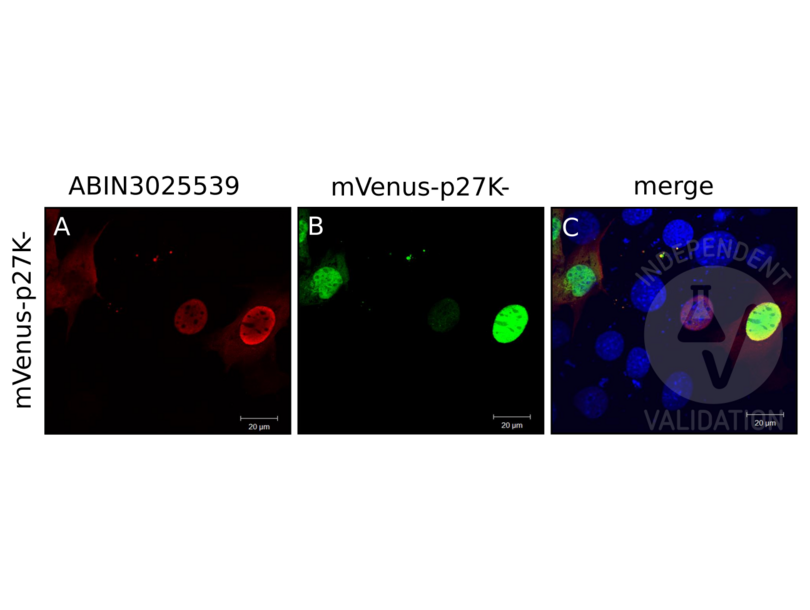
![NIH/3T3 cells were transfected with a plasmid encoding mVenus fused to p27K- and the expressed fusion protein detected by Venus autofluorescence (B, green) and by immunocytology using ABIN3025539 and anti-mouse AF555 (A, red). C shows red and green channels merged with DAPI counterstain (blue).]() NIH/3T3 cells were transfected with a plasmid encoding mVenus fused to p27K- and the expressed fusion protein detected by Venus autofluorescence (B, green) and by immunocytology using ABIN3025539 and anti-mouse AF555 (A, red). C shows red and green channels merged with DAPI counterstain (blue).
Full Methods
NIH/3T3 cells were transfected with a plasmid encoding mVenus fused to p27K- and the expressed fusion protein detected by Venus autofluorescence (B, green) and by immunocytology using ABIN3025539 and anti-mouse AF555 (A, red). C shows red and green channels merged with DAPI counterstain (blue).
Full Methods -
- 浓度
- 0.2 mg/mL
- 缓冲液
- 0.2 mg/mL in 1X PBS with 0.1 mg/mL BSA (US sourced) and 0.05 % sodium azide
- 储存液
- Sodium azide
- 注意事项
- This product contains Sodium azide: a POISONOUS AND HAZARDOUS SUBSTANCE which should be handled by trained staff only.
- 储存条件
- 4 °C,-20 °C
- 储存方法
- Store the p27 antibody cocktail at 2-8°C (with azide) or aliquot and store at -20°C or colder (without azide).
-
- 抗原
- P27
- 物质类
- Viral Protein
- 背景
- Recognizes a 27 kDa protein, identified as the p27Kip1, a cell cycle regulatory mitotic inhibitor. Its epitope spans between aa 83-204 of p27. It is highly specific and shows no cross-reaction with other related mitotic inhibitors. p27Kip1 functions as a negative regulator of G1 progression and has been proposed to function as a possible mediator of TGF- induced G1 arrest. p27Kip1 is a candidate tumor suppressor gene. This mAb co-precipitates cdk4 in complex p27Kip1 and is excellent for staining of formalin-fixed tissues.
-


