IL-6 Receptor 抗体 (PE-Cy7)
Quick Overview for IL-6 Receptor 抗体 (PE-Cy7) (ABIN2659081)
抗原
See all IL-6 Receptor (IL6R) 抗体适用
宿主
克隆类型
标记
应用范围
克隆位点
-
-
纯化方法
- The antibody was purified by affinity chromatography and conjugated with PE/Cy7 under optimal conditions. The solution is free of unconjugated PE/Cy7 and unconjugated antibody.
-
亚型
- IgG1 kappa
-
-
-
-
应用备注
- Optimal working dilution should be determined by the investigator.
-
限制
- 仅限研究用
-
-
- by
- Immunmodulatorische Abteilung, Universitätsklinikum Erlangen
- No.
- #101960
- 日期
- 2017.11.06
- 抗原
- IL6R
- Lot Number
- B236171
- Method validated
- Flow Cytometry
- Positive Control
- Mature dendritic cells (mDCs) from three donors, incubated with ABIN2659082
- Negative Control
- Mature dendritic cells (mDCs) from three donors, incubated without antibody
- Notes
Passed. ABIN2659082 specifically recognizes the IL-6 Receptor on mDCs.
- Primary Antibody
- ABIN2659082
- Secondary Antibody
- Full Protocol
- Prepare peripheral blood mononuclear cells (PBMCs) from leukoreduction system chambers (LRSCs) (Pfeiffer et al. (2013); Weidinger et al. 2011) from three healthy donors (obtained following informed consent and approved by the institutional review board) by density centrifugation on a Lymphoprep gradient (Axis-shield AS, 1114547, lot 12IFS10).
- For the generation of dendritic cells (DCs), prepare PBMCs from leukoreduction system chambers (LRSC) (Pfeiffer et al. (2013)).
- After centrifugation, collect PBMCs forming a distinct band at the sample/Lymphoprep interface.
- Wash PBMCs 3x with cold PBS supplemented with 1mM EDTA.
- Wash PBMCs 1x with cold RPMI 1640 (Lonza, BE12-167F, lot 7MB088).
- Resuspend PBMCs in warm DC Medium (RPMI 1640 supplemented with 1% AB-serum, 100U/ml penicillin, 100mg/ml streptomycin, 2mM L-glutamine, 10mM HEPES buffer (Lonza, BE17-737E)) and seeded into 175cm2 tissue culture flasks at a density of 3.5x10⁸ cells/flask.
- Let monocytes adhere for 1h at 37°C and 5% CO2.
- Wash non-adherent fraction off with warm RPMI 1640.
- Add 30ml DC medium supplemented with 800U/ml recombinant human granulocyte-macrophage colony-stimulating factor (GM-CSF) and 250U/ml recombinant human interleukin-4 (IL-4) to each flask (day 1).
- Incubate cells for 72h at 37°C and 5% CO2.
- On day 4 (or three days later), add 5ml of fresh culture medium containing GM-CSF (final concentration of 400 U/ml in 35ml) and IL-4 (final concentration of 250U/ml in 35ml) to the cells.
- On day 5, mature the immature DCs by adding 10ng/ml recombinant human tumor necrosis factor alpha (TNF-α), 1μg/ml prostaglandin E2 (PGE2), 200U/ml recombinant human interleukin-1β (IL-1β), 1000U/ml recombinant human interleukin-6 (IL-6), 40U/ml GM-CSF, and 250 U/ml IL-4 to the medium.
- Incubate cells for 48h at 37°C and 5% CO2.
- On day 7, use the now mature DCs (mDCs) for FACS analysis.
- Analyze cell surface expression of IL-6 Receptor by FACS:
- Harvest 2.5x10⁵ mDCs by centrifugation.
- Wash cells once with FACS buffer (PBS supplemented with 2% FCS).
- Combine anti-human PE-Cy7 conjugated IL-6 Receptor antibody (antibodies-online, ABIN2659082, lot B236171) diluted 1:100 and LIVE/DEAD Violet dead cell stain kit (ThermoFisher Scientific, L34955, lot 1832693) diluted 1:300 in cold FACS buffer.
- Resuspend mDCs in 100µl of the antibody-solution and incubate for 30min on ice in the dark.
- Wash mDCs 2x with cold FACS buffer.
- Fix mDCs using 2% paraformaldehyde in cold PBS.
- Store mDCs at 4°C until analysis.
- Run samples using a BD FACS Canto II and analyze events using FCS Express 5.
- Experimental Notes
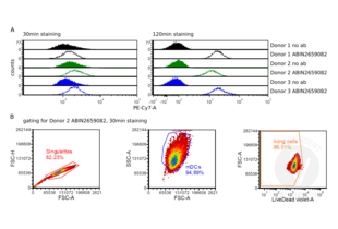
Mature DCs were stained for FACS for the analysis of the expression of IL-6 Receptor/CD126 on the cell surface of the cells with ABIN2659082 for 30min or 120min respectively. The histogram shows a clearly shift between unstained control and the stained mock control by staining for 120min. The staining for 30min does also show a shift of the histogram peak, but not as clear as the staining for 120min.
生效 #101960 (Flow Cytometry)![成功验证 '独立验证'标志]()
![成功验证 '独立验证'标志]() Validation Images
Validation Images![A Flow cytometry analysis of mature DCs from three different donors after incubation for 30min or 120min with or without ABIN2659082. B Consecutive gating for single cells (left panel), mDCs (middle panel), and living cells (right panel), illustrated on donor 2 samples stained for 30min with ABIN2659082.]() A Flow cytometry analysis of mature DCs from three different donors after incubation for 30min or 120min with or without ABIN2659082. B Consecutive gating for single cells (left panel), mDCs (middle panel), and living cells (right panel), illustrated on donor 2 samples stained for 30min with ABIN2659082.
Full Methods
A Flow cytometry analysis of mature DCs from three different donors after incubation for 30min or 120min with or without ABIN2659082. B Consecutive gating for single cells (left panel), mDCs (middle panel), and living cells (right panel), illustrated on donor 2 samples stained for 30min with ABIN2659082.
Full Methods -
-
缓冲液
- Phosphate-buffered solution, pH 7.2, containing 0.09 % sodium azide and 0.2 % (w/v) BSA .
-
储存液
- Sodium azide
-
注意事项
- This product contains Sodium azide: a POISONOUS AND HAZARDOUS SUBSTANCE which should be handled by trained staff only.
-
注意事项
- Protect from prolonged exposure to light. Do not freeze.
-
储存条件
- 4 °C
-
储存方法
- The antibody solution should be stored undiluted between 2°C and 8°C.
-
-
- IL-6 Receptor (IL6R) (Interleukin 6 Receptor (IL6R))
-
别名
- CD126
-
背景
- CD126 is an 80 kD IL-6 receptor α chain also known as IL-6R. It is a member of the immunoglobulin superfamily that is expressed on plasma cells, T cells, activated B cells, monocytes, granulocytes, hepatocytes, epithelial cells, and fibroblasts. Functional IL-6 receptors are formed by the non-covalent association of CD126 and the IL-6 receptor β chain (CD130 or gp130). CD126 binds IL-6 with low affinity but does not signal. The β chain (gp130, CD130) does not bind IL-6 by itself but associates with the α-chain/IL-6 complex to initiate signal transduction. IL-6 binding to the receptor complex results in the stimulation of B and T cells, and hematopoietic precursor proliferation and differentiation. A soluble form of CD126 has been found in human serum.
-
途径
- JAK/STAT Signaling, Autophagy, Growth Factor Binding, Cancer Immune Checkpoints
抗原
-


 (1 validation)


